Gel electrophoresis is a powerful technique used to separate and visualize DNA fragments based on their size. This method utilizes a three-dimensional gel matrix and an electrical current to facilitate the movement of DNA, which is inherently negatively charged. When DNA samples are loaded into wells at the top of the gel, they migrate towards the positively charged end, known as the anode, located at the bottom of the gel.
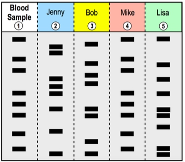
The separation of DNA occurs because larger fragments move more slowly through the gel matrix, remaining closer to their starting point at the top, while smaller fragments travel faster and thus migrate further down the gel. This size-based separation allows for the visualization of distinct bands representing different DNA fragments. Each lane of the gel typically contains a different DNA sample, with a reference lane that includes DNA fragments of known sizes, facilitating size comparison.
In a typical gel electrophoresis setup, the gel is organized into lanes, each containing wells where DNA samples are loaded. As the electrical current is applied, the negatively charged DNA moves towards the anode, creating a pattern of bands that can be analyzed. The distance traveled by each fragment correlates with its size; larger fragments travel shorter distances, while smaller fragments migrate further down the gel. This relationship can be graphically represented, showing that as the size of the DNA fragment decreases, the distance it travels increases.
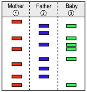
Understanding gel electrophoresis is crucial for various applications in molecular biology, including DNA fingerprinting, genetic analysis, and cloning. By comparing the bands of unknown samples to the reference DNA, researchers can estimate the sizes of the DNA fragments in the samples, providing valuable insights into genetic material.