Join thousands of students who trust us to help them ace their exams!Watch the first video
Multiple Choice
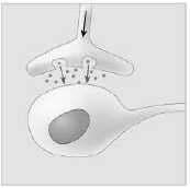
Which of the following types of signaling is represented in the figure?
A
Cell-cell recognition.
B
Paracrine.
C
Hormonal.
D
Synaptic.
0 Comments
Verified step by step guidance
1
Examine the image provided, which depicts two cells in close proximity with one cell releasing small molecules into the space between them.
Identify the structure in the image: the upper cell appears to be a neuron with a synaptic terminal, and the lower cell is likely another neuron or a target cell.
Observe the small dots between the two cells, which represent neurotransmitters being released from the synaptic terminal of the neuron.
Understand that synaptic signaling involves the release of neurotransmitters from a neuron into the synaptic cleft, which then bind to receptors on the adjacent cell.
Conclude that the image represents synaptic signaling, where communication occurs between neurons or between a neuron and a target cell through neurotransmitter release.
 Verified step by step guidance
 1:01m
1:01m