Gel electrophoresis is a powerful technique used to separate and visualize DNA fragments based on their size. This method utilizes a three-dimensional gel matrix and an electrical current to facilitate the movement of DNA, which is inherently negatively charged. When DNA samples are loaded into wells at the top of the gel, they migrate towards the positively charged end, known as the anode, located at the bottom of the gel.
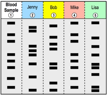
The separation of DNA occurs because larger fragments move more slowly through the gel matrix, remaining closer to their starting point at the top, while smaller fragments travel faster and thus migrate further down the gel. This size-based separation allows for the visualization of distinct bands representing different DNA fragments. Each lane of the gel typically contains a different DNA sample, with a reference lane that includes DNA fragments of known sizes, measured in base pairs (bp).
As the DNA migrates, the relationship between fragment size and distance traveled can be graphically represented. A graph plotting molecular size against migration distance shows that larger DNA fragments travel shorter distances, while smaller fragments cover more ground. This principle is crucial for analyzing DNA samples, as it enables comparison against the reference lane to estimate the sizes of unknown fragments.
In summary, gel electrophoresis is an essential technique in molecular biology for separating DNA fragments by size, allowing researchers to visualize and analyze genetic material effectively. Understanding the mechanics of this process is fundamental for applications in genetics, forensic science, and biotechnology.