Pearson+ LogoPearson+ Logo
Ch. 3 - Cell Division and Chromosome Heredity
Sanders - Genetic Analysis: An Integrated Approach 3rd Edition
Sanders3rd EditionGenetic Analysis: An Integrated ApproachISBN: 9780135564172Not the one you use?Change textbook
Chapter 3, Problem C.3c

For the retinal cancer retinoblastoma, the inheritance of one mutated copy of RB1 from one of the parents is often referred to as a mutation that produces a 'dominant predisposition to cancer.' This means that the first mutation does not produce cancer but makes it very likely that cancer will develop.
Using RB1⁺ for the normal wild-type allele and RB1⁻ for the mutant allele, identify the genotype of a cell in a retinoblastoma tumor.

Verified step by step guidance
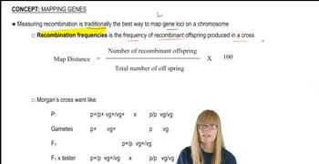
1
Understand that retinoblastoma is caused by mutations in the RB1 gene, where RB1⁺ represents the normal (wild-type) allele and RB1⁻ represents the mutant allele.
Recognize that individuals with a dominant predisposition inherit one mutated allele (RB1⁻) but cancer develops only after a second mutation occurs in the other allele within a cell.
Recall the 'two-hit hypothesis' which states that both copies of the RB1 gene must be inactivated (mutated) in a single cell for the tumor to form.
Therefore, the genotype of a cell in a retinoblastoma tumor will have both alleles mutated, meaning it will be homozygous mutant: RB1RB1.
Summarize that the tumor cell genotype is RB1^{-}RB1^{-}, indicating loss of function of both RB1 alleles, which leads to uncontrolled cell growth and cancer.

Verified video answer for a similar problem:

This video solution was recommended by our tutors as helpful for the problem above.
Video duration:
1m
Was this helpful?

Key Concepts

Here are the essential concepts you must grasp in order to answer the question correctly.

Tumor Suppressor Genes and the RB1 Gene

Tumor suppressor genes like RB1 produce proteins that regulate cell growth and prevent cancer. The RB1 gene encodes a protein that controls the cell cycle, and mutations in RB1 can lead to loss of this control, promoting tumor development. Both copies of the gene typically need to be inactivated for cancer to occur.
Recommended video:
Guided course
09:09
Mapping Genes

Knudson's Two-Hit Hypothesis

This hypothesis explains that two mutations ('hits') are required to inactivate both alleles of a tumor suppressor gene for cancer to develop. In hereditary retinoblastoma, one mutated allele (RB1⁻) is inherited, and a second somatic mutation inactivates the other allele, leading to tumor formation.
Recommended video:
Guided course
03:13
Translation:Wobble Hypothesis

Genotype of Retinoblastoma Tumor Cells

In retinoblastoma tumor cells, both RB1 alleles are mutated or inactivated (RB1⁻/RB1⁻), resulting in loss of tumor suppressor function. Although the inherited mutation is dominant in predisposition, the tumor cells themselves are homozygous or functionally null for RB1, driving cancer progression.
Recommended video:
Guided course
03:52
Cell-cell interactions
Related Practice
Textbook Question

For the retinal cancer retinoblastoma, the inheritance of one mutated copy of RB1 from one of the parents is often referred to as a mutation that produces a 'dominant predisposition to cancer.' This means that the first mutation does not produce cancer but makes it very likely that cancer will develop.


What is the genotype of a normal cell in the retina in a person who has sporadic retinoblastoma? What is the normal cell genotype if the person has hereditary retinoblastoma? Explain the reason for the difference between the genotypes.

3
views
Textbook Question

From a piece of blank paper, cut out three sets of four cigar-shaped structures (a total of 12 structures). These will represent chromatids. Be sure each member of a set of four chromatids has the same length and girth. In set one, label two chromatids 'A' and two chromatids 'a.' Cut each of these chromatids about halfway across near their midpoint and slide the two 'A' chromatids together at the cuts, to form a single set of attached sister chromatids. Do the same for the 'a' chromatids. In the second set of four chromatids, label two 'B' and two 'b.' Cut and slide these together as you did for the first set, joining the 'B' chromatids together and the 'b' chromatids together. Repeat this process for the third set of chromatids, labeling them as 'D' and 'd.' You now have models for three pairs of homologous chromosomes, for a total of six chromosomes. Align the chromosomes as they might appear at metaphase I of meiosis.

1
views
Textbook Question

Examine the following diagrams of cells from an organism with diploid number 2n=6, and identify what stage of M phase is represented.

1
views