Pearson+ LogoPearson+ Logo
Ch. 10 - Eukaryotic Chromosome Abnormalities and Molecular Organization
Sanders - Genetic Analysis: An Integrated Approach 3rd Edition
Sanders3rd EditionGenetic Analysis: An Integrated ApproachISBN: 9780135564172Not the one you use?Change textbook
Chapter 10, Problem 27a

Genomic DNA from the nematode worm Caenorhabditis elegans is organized by nucleosomes in the manner typical of eukaryotic genomes, with 145 bp encircling each nucleosome and approximately 55 bp in linker DNA. When C. elegans chromatin is carefully isolated, stripped of nonhistone proteins, and placed in an appropriate buffer, the chromatin decondenses to the 10-nm fiber structure. Suppose researchers mix a sample of 10-nm–fiber chromatin with a large amount of the enzyme DNase I that randomly cleaves DNA in regions not protected by bound protein. Next, they remove the nucleosomes, separate the DNA fragments by gel electrophoresis, and stain all the DNA fragments in the gel.
Approximately what range of DNA fragment sizes do you expect to see in the stained electrophoresis gel? How many bands will be visible on the gel?

Verified step by step guidance
1
Step 1: Understand the structure of chromatin in Caenorhabditis elegans. Chromatin is organized into nucleosomes, where 145 base pairs (bp) of DNA are wrapped around histone proteins, and approximately 55 bp of linker DNA connects adjacent nucleosomes. This organization is typical of eukaryotic genomes.
Step 2: Recognize the role of DNase I in the experiment. DNase I is an enzyme that randomly cleaves DNA in regions not protected by bound proteins, such as the linker DNA between nucleosomes. The nucleosomal DNA (145 bp) is protected from cleavage due to its association with histones.
Step 3: Predict the DNA fragment sizes after DNase I treatment. Since DNase I cleaves the linker DNA (approximately 55 bp), the resulting DNA fragments will consist of the protected nucleosomal DNA (145 bp) plus varying amounts of linker DNA. The smallest fragment size will be approximately 145 bp, and the largest fragment size will be 145 bp + 55 bp = 200 bp.
Step 4: Consider the separation of DNA fragments by gel electrophoresis. Gel electrophoresis separates DNA fragments based on size. Since the DNA fragments range from 145 bp to 200 bp, you would expect to see distinct bands corresponding to these sizes, depending on the resolution of the gel.
Step 5: Determine the number of bands visible on the gel. The number of bands will depend on the variability in linker DNA cleavage by DNase I. If DNase I cleaves at random positions within the linker DNA, you may see multiple bands corresponding to different fragment sizes between 145 bp and 200 bp. However, if cleavage is consistent, fewer distinct bands may be visible.

Verified video answer for a similar problem:

This video solution was recommended by our tutors as helpful for the problem above.
Video duration:
4m
Was this helpful?

Key Concepts

Here are the essential concepts you must grasp in order to answer the question correctly.

Nucleosome Structure

Nucleosomes are the fundamental units of chromatin, consisting of a segment of DNA wrapped around a core of histone proteins. Each nucleosome typically contains about 145 base pairs (bp) of DNA, which is crucial for the compaction and organization of eukaryotic genomes. Understanding nucleosome structure is essential for predicting how DNA is protected from enzymatic cleavage and how it contributes to the overall architecture of chromatin.
Recommended video:
Guided course
07:10
Chromosome Structure

DNase I Activity

DNase I is an enzyme that cleaves DNA at sites that are not protected by bound proteins, such as histones in nucleosomes. When chromatin is treated with DNase I, the enzyme will cut the linker DNA and any exposed regions, resulting in fragments of varying sizes. This concept is vital for interpreting the results of gel electrophoresis, as the size of the resulting DNA fragments will depend on the accessibility of the DNA to the enzyme.
Recommended video:
Guided course
08:04
Decision Between Lytic and Lysogenic Cycles

Gel Electrophoresis

Gel electrophoresis is a laboratory technique used to separate DNA fragments based on their size. In this process, DNA samples are loaded into a gel matrix and subjected to an electric field, causing smaller fragments to migrate faster than larger ones. The resulting pattern of bands on the gel provides insight into the sizes of the DNA fragments generated by DNase I digestion, allowing researchers to estimate the number of bands and their corresponding sizes.
Recommended video:
Related Practice
Textbook Question

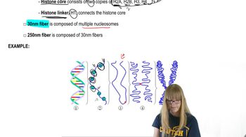
Experimental evidence demonstrates that the nucleosomes present in a cell after the completion of S phase are composed of some 'old' histone dimers and some newly synthesized histone dimers. Describe the general design for an experiment that uses a protein label such as ³⁵S to show that nucleosomes are often a mixture of old and new histone dimers following DNA replication.

Textbook Question

DNase I cuts DNA that is not protected by bound proteins but is unable to cut DNA that is complexed with proteins. Human DNA is isolated, stripped of its nonhistone proteins, and mixed with DNase I. Samples are removed after 30 minutes, 1 hour, and 4 hours and run separately in gel electrophoresis. The resulting gel is stained to make all DNA fragments in it visible, and the results are shown in the figure. DNA fragment sizes in base pairs (bp) are estimated by the scale to the left of the gel. Examine the gel results and speculate why longer DNase I treatment produces different results.

4
views
Textbook Question

DNase I cuts DNA that is not protected by bound proteins but is unable to cut DNA that is complexed with proteins. Human DNA is isolated, stripped of its nonhistone proteins, and mixed with DNase I. Samples are removed after 30 minutes, 1 hour, and 4 hours and run separately in gel electrophoresis. The resulting gel is stained to make all DNA fragments in it visible, and the results are shown in the figure. DNA fragment sizes in base pairs (bp) are estimated by the scale to the left of the gel. Draw a conclusion about the organization of chromatin in the human genome from this gel.

Textbook Question

Genomic DNA from the nematode worm Caenorhabditis elegans is organized by nucleosomes in the manner typical of eukaryotic genomes, with 145 bp encircling each nucleosome and approximately 55 bp in linker DNA. When C. elegans chromatin is carefully isolated, stripped of nonhistone proteins, and placed in an appropriate buffer, the chromatin decondenses to the 10-nm fiber structure. Suppose researchers mix a sample of 10-nm–fiber chromatin with a large amount of the enzyme DNase I that randomly cleaves DNA in regions not protected by bound protein. Next, they remove the nucleosomes, separate the DNA fragments by gel electrophoresis, and stain all the DNA fragments in the gel.

Explain the origin of DNA fragments seen in the gel.

3
views
Textbook Question

Genomic DNA from the nematode worm Caenorhabditis elegans is organized by nucleosomes in the manner typical of eukaryotic genomes, with 145 bp encircling each nucleosome and approximately 55 bp in linker DNA. When C. elegans chromatin is carefully isolated, stripped of nonhistone proteins, and placed in an appropriate buffer, the chromatin decondenses to the 10-nm fiber structure. Suppose researchers mix a sample of 10-nm–fiber chromatin with a large amount of the enzyme DNase I that randomly cleaves DNA in regions not protected by bound protein. Next, they remove the nucleosomes, separate the DNA fragments by gel electrophoresis, and stain all the DNA fragments in the gel.

How do the expected results support the 10-nm–fiber model of chromatin?

Textbook Question

A small population of deer living on an isolated island is separated for many generations from a mainland deer population. The populations retain the same number of chromosomes but hybrids are infertile. One chromosome (shown here) has a different banding pattern in the island population than in the mainland population.

Describe how the banding pattern of the island population chromosome most likely evolved from the mainland chromosome. What term or terms describe the difference between these chromosomes?